In a lab in Olin Science Center, a high-definition monitor displays a yellow oval object with two vertical blue lines.
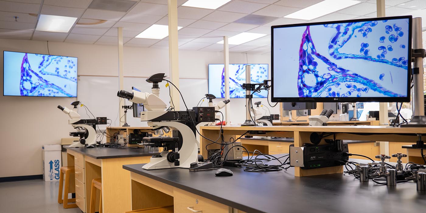
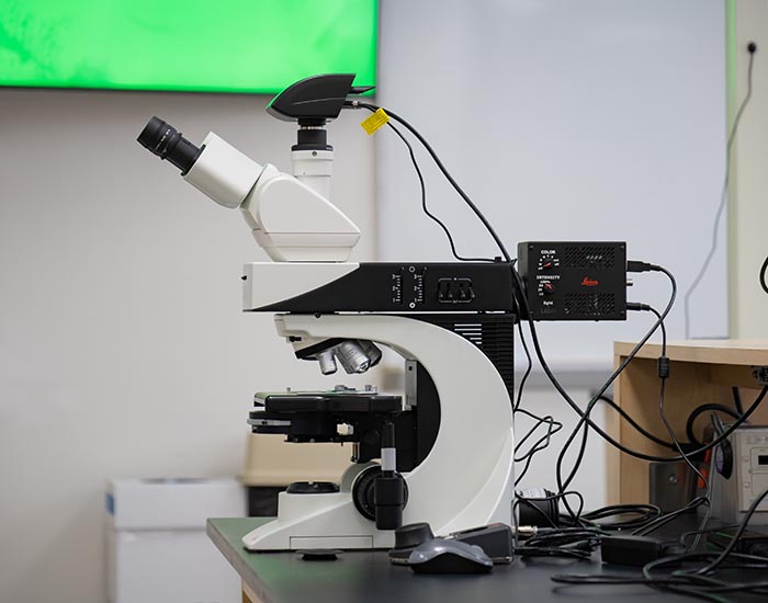
What at first appears to be a magnified pill is the embryo of a fruit fly, only a few hours old — the blue lines highlight the distinct pattern of a gene’s expression. The embryo, carefully stained and mounted on a slide by biology major Eleanor McDonough ’22, is being projected from a Leica DM2500 compound microscope, one of several within a new microscopy classroom in the Department of Biology.
Viewing such a tiny object in this manner with the department’s former equipment was unthinkable. But the new suite of state-of-the-art microscopes is making the impossible possible for all Willamette University students, who will have the ability to perform graduate-level research and examine specimens in a variety of ways starting this fall.
The department bought the microscopes — and plans to purchase another set of complementary microscopes — to make the classroom among the first of its kind, and an exceptional learning environment for students from a breadth of disciplinary studies, said Associate Professor of Biology Jason Duncan.
“The classroom opens up a whole new world for students,” he said. “They will be able to see the microscopic world in ways they never imagined, and do so in a very collaborative and engaging way.”
Building on a foundational skill
Microscopy is a foundational skill in biology and critical for fields including botany, anatomy and medicine.
For science majors, the classroom’s capabilities will strengthen their appeal to future employers as workforce demand for this training is high. But even first-year students with a vague interest in science will find it hard not to be impressed. “We hope that students who haven’t even considered biology will be so excited by what they see in this space that they’ll reconsider,” Duncan said.
Cells invisible to the human eye can be observed in exquisite detail with the new microscopes, which are also equipped with special filters to provide contrast or enhance or illuminate a specimen — students can tinker with it in the same way they might edit an image in Adobe Photoshop.
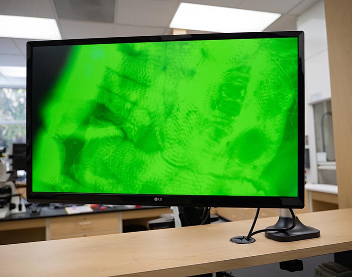
One of the microscope’s settings, epifluorescence, allows students to observe structures within a cell. Scientists have engineered proteins to emit fluorescent light, generating an array of colors that can be used to highlight structures within the tiniest cells and organisms. Under the right combination of wavelengths, fibroblast cells — the long, thin cells responsible for the stretchiness in skin — reveal glowing blue nuclei, red mitochondria and green microfilaments.
Potential for collaboration across campus
The power of the new equipment is not only what students can see, but how they see it.
Sudden observations can be immediately shared with the entire class, and a 4K ultra high-definition digital camera attached to each microscope allows students to shoot photos and video. Although the Leicas can’t fully capture 3D or extra large specimens, the biology department plans on upgrading with software to create composite images of larger specimens in their entirety.
The classroom won’t be limited to science majors, either. First-year students in introductory biology courses will have access to the classroom, and the department wants to open it to other groups across campus — exercise and health science students might use the microscopes to study gross anatomy, such as cross-sections of muscle or other tissues, while art majors could use the space to create artistic images of biological materials, said Duncan.
Microscopy sits at the intersection of many disciplines. Understanding the optics of light and how it interacts with a specimen touches on physics, while observing and imaging specimens stained with fluorescent dyes requires a knowledge of chemistry. Analyzing a digital image, composed of millions of pixels, requires a grounding in data science.
“Viewing the world through a microscope is also a pretty cool way to explore it,” stated Duncan.


